
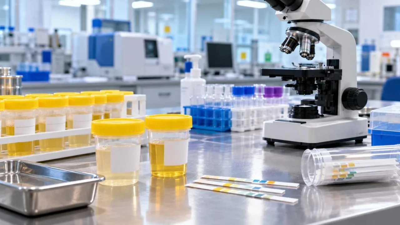
The urine routine and microscopy test is quick, cheap and surprisingly informative. It can point to urinary infections, kidney disease, diabetes, dehydration and even liver problems. Here is how to make sense of the report.
How to give a good sample
- Use the sterile container from the lab and do not touch the inside.
- Wash the genital area with water first.
- Collect a midstream sample: pass the first part of the urine into the toilet, then collect the middle portion.
- A first-morning sample is often preferred, and it should reach the lab within an hour or two.
- Women should ideally avoid testing during menstruation, as blood can contaminate the sample.
Physical examination
| Parameter | Usual finding | What changes may mean |
|---|---|---|
| Colour | Pale to dark yellow | Very dark: dehydration or bilirubin; red or brown: blood, certain foods or medicines |
| Appearance | Clear | Cloudy: infection, crystals or mucus |
| Specific gravity | about 1.005 – 1.030 | High: dehydration; persistently low: kidney concentrating problems |
| pH | about 4.5 – 8.0 | Can be influenced by diet, infection and stone risk |
Chemical examination
- Protein (albumin): normally nil or trace. Persistent protein can signal kidney damage, often from diabetes or high blood pressure. Fever, heavy exercise and standing for long periods can cause temporary traces.
- Glucose (sugar): normally nil. Sugar in urine usually means blood sugar is high, and diabetes should be checked.
- Ketones: normally nil. They appear with fasting, very low-carb diets, vomiting or, more seriously, uncontrolled diabetes.
- Bilirubin and urobilinogen: changes can point to liver or bile duct problems.
- Nitrite and leukocyte esterase: positive results suggest a bacterial urinary tract infection.
- Blood: normally negative; see the RBC count below.
Microscopic examination
| Finding | Usual range | Possible meaning if raised |
|---|---|---|
| Pus cells (WBCs) | 0 – 5 per high-power field | Urinary tract infection or inflammation |
| Red blood cells | 0 – 2 per high-power field | Stones, infection, injury, kidney disease; needs follow-up |
| Epithelial cells | Few | Many: often a contaminated sample |
| Casts | None or occasional hyaline | Some types suggest kidney disease |
| Crystals | Occasional | Many: stone risk, diet or dehydration |
| Bacteria | Nil or few | Infection or contamination |
Urinary tract infection (UTI) clues
Burning while passing urine, frequent urge, lower abdominal pain or fever, together with raised pus cells and positive nitrite, suggest a UTI. Your doctor may order a urine culture to identify the bacteria and choose the right antibiotic. Avoid taking leftover antibiotics on your own, which can fuel resistance.
When to see a doctor
Consult a doctor if your report shows protein, sugar, blood or many pus cells, or if you have pain, fever, blood in the urine, swelling of the feet or face, or a sudden change in how much you urinate.
Lab Tests Explained: the full series
- Part 1: CBC (Complete Blood Count)
- Part 2: Lipid Profile
- Part 3: Liver Function Test
- Part 4: Kidney Function Test
- Part 5: Urine Routine Test (you are here)
- Part 6: Iron Studies and Ferritin
- Part 7: Vitamin B12
- Part 8: Full-Body Checkup by Age
Related reads: Blood sugar levels and HbA1c · Thyroid tests · Vitamin D test · Uric acid
Disclaimer: This article is for general information only and is not medical advice. Reference ranges vary between laboratories, so always read your report against the range printed on it and discuss results with a qualified doctor.